Brain region generalisation
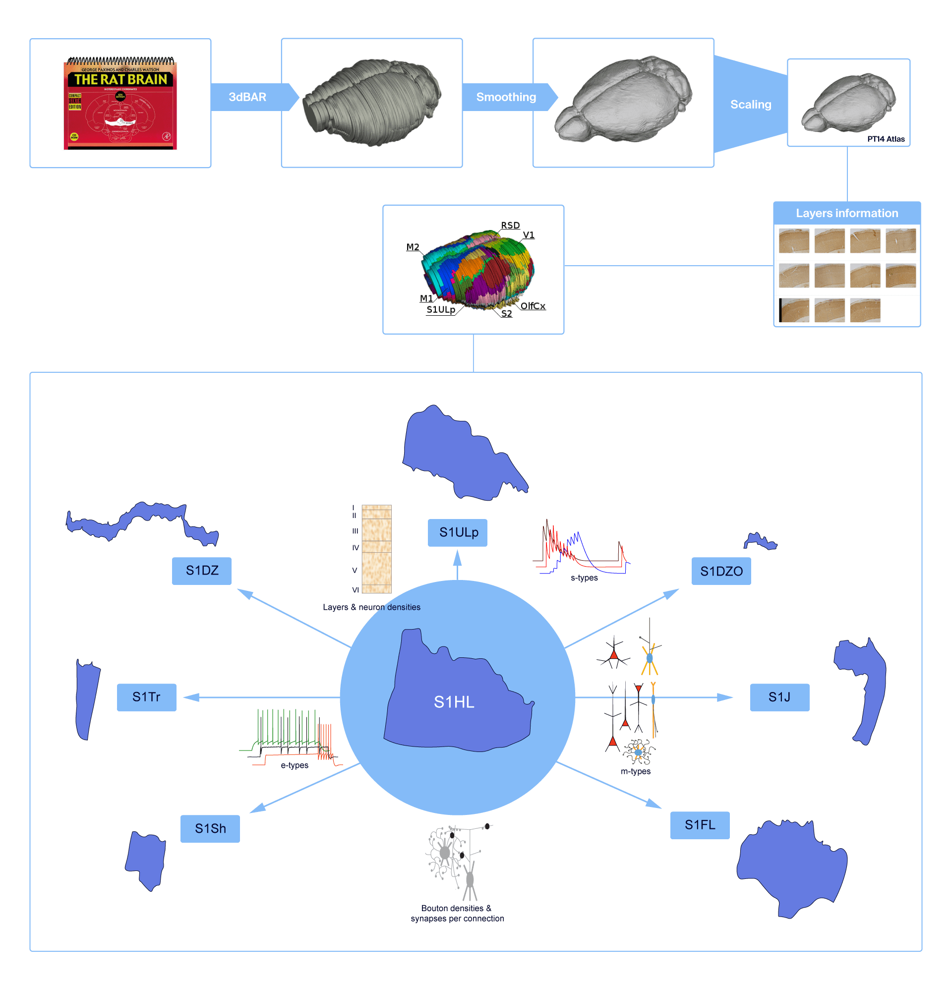
The infographic depicts how sparse experimental data was extrapolated to yield a dense, multi-scale reconstruction of 8 sub-regions of the rat primary somatosensory cortex.
We procured the following sparse experimental data from the hind limb representation of juvenile rat primary somatosensory cortex (S1 HL):
- Layer heights
- Neuron densities
- Synapse densities
- Morphological reconstructions
- Layer-wise proportions of morphological types (m-types)
- Electrophysiological recordings (e-types)
- Morpho-electrical proportions (me-types)
- M-type specific bouton densities
- No. of synapses/connection and patterns of their axo-dendritic innervation
- synapse types (s-types) based on release probability, time constants for recovery from depression/facilitation, quantal conductances, and reversal potentials recorded at 2 mM [Ca2+]o
We then extrapolated these data to 7 other sub-regions of rat S1: S1FL, "Fore Limb", S1Sh, "Shoulder", S1Tr, "Trunk", S1J, “Jaw", S1ULp, "Upper lip", S1DZ, "Disgranular zone", S1DZO, "Oral disgranular zone" to yield a dense, multi-scale reconstruction of the entire S1.